A clinical, evidence-based guide for chiropractors, physical therapists, and primary care physicians.
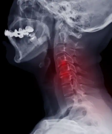
Cervical spinal cord compression is one of the most time-sensitive conditions in spine care. Many degenerative cervical spine conditions can be managed conservatively, but cervical cord compression with neurological impairment often requires surgery to prevent irreversible injury. The clinical challenge is separating radiographic cord compression, which is common in aging populations, from symptomatic cervical myelopathy, which carries a significantly higher risk of progressive and permanent disability if left untreated.
What Causes Cervical Cord Compression?
Cord compression most commonly results from degenerative changes collectively referred to as cervical spondylotic myelopathy (CSM), the leading cause of spinal cord dysfunction in adults over age 55.
Mechanisms of compression include:
These processes reduce the space available for the spinal cord, resulting in direct mechanical compression, vascular compromise, impaired axonal conduction, and progressive neuronal injury. Chronic compression leads to demyelination, axonal loss, and spinal cord atrophy. Because the spinal cord has limited capacity for regeneration, early identification and treatment are critical.
When Does Cord Compression Become Myelopathy?
Not all cord compression requires surgery. The key determinant is the presence of clinical myelopathy, which reflects spinal cord dysfunction rather than an incidental imaging finding.
Early Symptoms of Cervical Myelopathy
Patients often present with subtle, progressive symptoms, including hand clumsiness, difficulty with fine motor tasks, loss of dexterity, hand numbness, balance impairment, and gait instability. Practically, this shows up as trouble buttoning shirts, writing, using keys, or opening containers. These symptoms are frequently mistaken for peripheral nerve disorders, which is why recognizing the early signs of cervical myelopathy matters at the frontline.
Objective Neurological Findings
Objective examination findings are the most important indicators of clinically significant cord compression.
- Upper motor neuron signs: hyperreflexia, Hoffmann sign, Babinski sign, clonus, spasticity, increased muscle tone
- Motor findings: intrinsic hand weakness, grip weakness, loss of coordination
- Gait findings: wide-based gait, unsteady gait, reduced stride length
These findings reflect spinal cord dysfunction rather than isolated nerve root pathology, and they carry more weight in referral decisions than imaging alone.
Have a patient with signs of myelopathy?
Dr. Wascher offers a free MRI review and second opinion for referring providers, with documented career outcomes available for full transparency. Fast surgical evaluation when it counts.
Call (855) 854-9274 or request a consultation for your patient.
Surgical Indications: What Tips the Balance Toward Surgery
Progressive Neurological Deficit
Progressive neurological decline is one of the strongest indications for surgery. Examples include worsening hand weakness, increasing gait instability, declining coordination, and progressive loss of dexterity. Progressive myelopathy rarely improves without surgical intervention, and neurological deterioration often continues while compression persists. Published natural-history data indicate that roughly 20% to 60% of patients with symptomatic myelopathy deteriorate neurologically over time without surgery. Earlier surgical intervention improves the likelihood of neurological recovery.
Functional Decline and Disability
Functional impairment is a key determinant in surgical decision-making. Indicators include loss of independence, difficulty walking safely, frequent falls, impaired hand function, and reduced ability to perform daily activities. Functional decline reflects clinically significant spinal cord dysfunction, and surgery is often recommended to prevent further deterioration.
MRI Findings That Support Surgical Referral
MRI is the gold standard for evaluating cervical cord compression. For a fuller breakdown, see our guide to MRI findings that warrant referral for cervical spine disease. Concerning findings include the following.
Cord Compression With Clinical Myelopathy
This combination is the single most important surgical indication. Findings may include cord flattening, cord indentation, and loss of cerebrospinal fluid around the cord.
Spinal Cord Signal Change
T2 hyperintensity within the spinal cord indicates cord injury, reflecting edema, gliosis, or myelomalacia. Signal change correlates with more severe neurological injury, an increased likelihood of progression, and worse outcomes without treatment. Patients with cord signal change and symptoms should be referred promptly.
Severe Canal Stenosis
Severe narrowing increases the risk of neurological deterioration. A canal diameter below approximately 10 mm is generally considered severe stenosis and increases vulnerability to spinal cord injury. Patients with severe cervical stenosis warrant closer evaluation.
Failure of Conservative Management
Conservative care may be appropriate for patients with mild symptoms, no neurological deficits, and a stable condition. Surgery becomes appropriate when neurological deficits develop, symptoms progress, or functional decline occurs. Physical therapy and conservative care do not decompress the spinal cord. Their role is supportive, not curative, in cases of myelopathy.
Natural History and the Risk of Waiting
The natural history of untreated cervical myelopathy is typically progressive. Neurological decline may be gradual or stepwise, and some patients experience sudden deterioration. Once spinal cord injury progresses, recovery becomes less predictable, and delayed surgery is associated with worse outcomes.
Patients with cord compression are also at increased risk of acute neurological worsening following falls, minor trauma, or sudden neck movement. Severe stenosis reduces the protective reserve around the cord, so even minor trauma may cause significant injury. This is a key reason to identify at-risk patients early.
Surgical Goals and Expected Outcomes
The primary goal of surgery is to decompress the spinal cord and prevent further neurological deterioration. Secondary goals include improving neurological function, gait and balance, and hand function, and reducing disability. Surgical approaches may include anterior cervical decompression, posterior cervical decompression, or corpectomy, often with intraoperative neuromonitoring to protect cord function. Surgery is more effective at halting progression than reversing severe deficits, so earlier surgery generally produces better outcomes.
When Should a Provider Refer?
Prompt referral is appropriate when any of the following are present:
- Objective signs of myelopathy
- Progressive neurological deficit
- Cord signal change on MRI
- Functional decline
- Severe cord compression with symptoms
Delayed referral may result in permanent neurological impairment. Cord compression without symptoms does not always require surgery, but these patients should be monitored closely, and referral may still be appropriate for severe stenosis, cord signal change, or high-risk anatomy. Early evaluation allows appropriate monitoring and planning.
Chiropractors, physical therapists, and primary care physicians play a critical role in early detection through careful neurological examination, recognition of upper motor neuron signs, monitoring of symptom progression, appropriate imaging, and timely referral. When distinguishing myelopathy from mimics, keep peripheral neuropathy, carpal tunnel syndrome, ulnar neuropathy, multiple sclerosis, and motor neuron disease in the differential. Neurological examination and MRI help separate these conditions.
Key Surgical Indications at a Glance
Surgery is generally indicated when cervical cord compression is associated with:
- Clinical myelopathy
- Progressive neurological deficit
- Functional impairment
- Cord signal change on MRI
- Severe cord compression with symptoms
Refer with confidence
When myelopathy is on the table, timing changes outcomes. Dr. Thomas Wascher has performed more than 4,500 cervical spine surgeries and publishes his career outcomes for every case. Send your patient for a fast, transparent surgical evaluation.
Call (855) 854-9274 or request a free MRI review. See our documented outcomes.
Frequently Asked Questions
When does cervical cord compression require surgery?
Surgery is generally indicated when cord compression is accompanied by clinical myelopathy, a progressive neurological deficit, functional decline, cord signal change on MRI, or severe compression with symptoms. Radiographic compression alone, without symptoms or exam findings, does not automatically require surgery.
Does cord compression seen on MRI always require surgery?
No. Radiographic cord compression is common in aging spines and is not, by itself, a surgical indication. The presence of clinical myelopathy or a progressive deficit is what tips the balance toward surgical referral.
What early signs of cervical myelopathy should frontline providers look for?
Watch for hand clumsiness, loss of dexterity, difficulty with fine motor tasks, gait instability, and balance problems, along with upper motor neuron signs such as hyperreflexia, a positive Hoffmann sign, or clonus. These are often mistaken for peripheral nerve conditions.
How urgently should a patient with suspected myelopathy be referred?
Promptly. Progressive myelopathy rarely improves without surgery, and delayed treatment is associated with worse outcomes. Objective signs of myelopathy or a progressive deficit warrant timely surgical evaluation rather than prolonged conservative care.
Is monitoring appropriate for asymptomatic cord compression?
Cord compression without symptoms does not always require surgery, but these patients should be monitored closely. Referral may still be appropriate for severe stenosis, cord signal change on MRI, or high-risk anatomy, so early evaluation supports better planning.
Authored and reviewed by Thomas Wascher, MD, FACS, of Wascher Cervical Spine Institute in Appleton, WI. This article is intended as clinical reference material for referring providers and does not replace individualized medical judgment.