Whether you're wondering about your options or ready to schedule a consultation, we're here to help you take the next step toward a pain-free life.

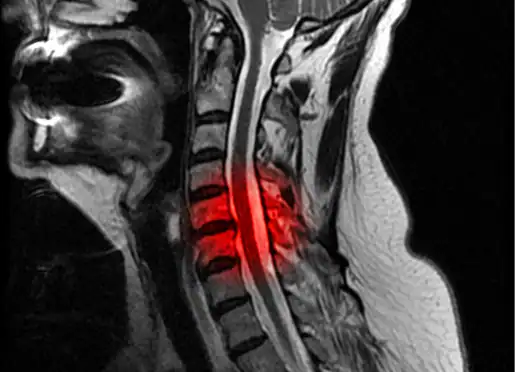
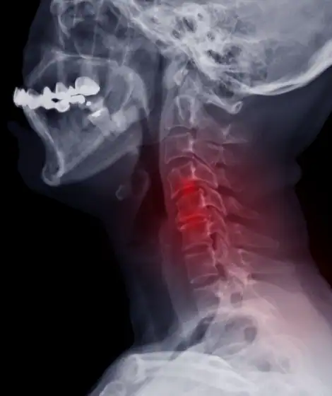
Ossification of the posterior longitudinal ligament (OPLL), commonly called OPLL, is a rare condition in which a spinal ligament in your neck abnormally hardens into bone, compressing the spinal cord and causing progressive myelopathy. This ligament normally runs behind your vertebrae from the skull to the lower spine, helping limit excessive neck flexion. In OPLL, abnormal bone formation fuses the ligament and dura into a solid sheet of bone in front of the spinal cord, making surgical decompression exceptionally challenging. OPLL is most commonly seen in patients of Asian descent, affecting approximately 4-5% of the Japanese population and 1.5% of the U.S. population. Most patients become symptomatic between the ages of 40 and 70, presenting with progressive difficulty walking, hand coordination problems, burning arm pain, and upper extremity weakness and numbness. At Wascher Cervical Spine Institute in Appleton, WI, Dr. Thomas Wascher has achieved a 99% improvement rate, treating over 252+ cases using advanced microsurgical techniques.
The exact cause of ossification of the posterior longitudinal ligament remains poorly understood, though research points to both genetic and environmental factors. The condition involves extensive abnormal bone formation in the posterior longitudinal ligament, a structure that extends from the base of the skull to the sacrum, located immediately behind the vertebral bodies and discs.
In OPLL, the ossification process often involves both the ligament and the adjacent dura concurrently, causing the dura to become incorporated into the resulting sheet of bone anterior to the spinal cord. This creates particular challenges for surgical treatment, as resection of the calcified mass requires resection of the dura as well, creating a very high risk of cerebrospinal fluid leak and potential spinal cord injury.
OPLL is more common in the cervical spine than in the thoracic or lumbar segments, although the entire spine may be involved in some cases.
Two forms of cervical OPLL have been identified:
Continuous OPLL: A longitudinal sheet of bone extending behind the vertebral bodies. This type is more likely to result in eventual myelopathy and typically requires surgical intervention.
Segmental OPLL: Focal calcification primarily at the level of the disc spaces. While less likely to cause immediate symptoms, patients with segmental OPLL still require monitoring and are at increased risk for spinal cord injury from minor falls.

Common Symptoms: OPLL myelopathy typically presents with progressive symptoms including:
Even asymptomatic patients with radiographic evidence of OPLL are at increased risk for spinal cord injury from minimal trauma or falls.
Diagnostic Process: OPLL diagnosis requires both MRI and CT imaging of the cervical spine:
MRI: Determines the degree of spinal cord compression and identifies the number of affected levels. May reveal signal changes within the spinal cord indicating myelomalacia.
CT Scan: Confirms the presence of abnormal bone anterior to the spinal cord and measures the critical occupation ratio.
Occupation Ratio: A measure of the anterior-posterior distance of the spinal canal occupied by abnormal bone divided by the expected normal anterior-posterior distance at that level. This ratio predicts disease severity:
Conservative Management: OPLL treatment begins with conservative management for patients with radiographic evidence of OPLL but no overt myelopathy, involving close monitoring. However, progression is common once myelopathic symptoms appear, and operative decompression becomes necessary.
Surgical Approaches for OPLL: OPLL surgery is technically demanding due to the fusion of bone and dura. Three main approaches exist:
Anterior Decompression (Anterior Cervical Corpectomy): For severe cases, multilevel anterior cervical corpectomy using structural allograft and anterior plating provides direct decompression. This approach offers the greatest rate of neurologic recovery but requires extreme care and precise microsurgical techniques to avoid cerebrospinal fluid leak. A floating decompression technique may be used, leaving a thin island of bone (2-3 mm) composed of OPLL remnants that floats anteriorly, minimizing CSF leak risk.
Posterior Decompression (Posterior Cervical Decompression and Fusion): Laminectomies with fusion and lateral mass or pedicle screw instrumentation. This approach is avoided as the sole treatment in patients with significant cervical kyphosis (greater than 15-20 degrees). Laminoplasty is not recommended due to unacceptably high rates of chronic pain, post-operative C5 radiculopathy, and recurrent stenosis.
Combined Anterior-Posterior Approach (Preferred for Severe Cases): For patients with an occupation ratio of 60% or greater, combined decompression yields the best results. Dr. Wascher's preference is to perform multilevel anterior corpectomy as Stage I, followed within days to weeks by posterior laminectomies with fusion and instrumentation. This staged approach increases fusion rates, recreates the normal posterior cervical tether, and prevents post-operative kyphosis.
Intraoperative neuromonitoring is essential during OPLL surgery to continuously monitor spinal cord function and minimize risk.
Vanessa had years of neck pain leaving her unable to even do her daily work. But with Dr. Wascher’s quick and timely intervention that included multiple viewings of MRIs, muscle and nerve tests, followed by a 3-Level Anterior Cervical Fusion, she is now happy without any neck issues. “I can happily say that by following the recommendations of Dr. Wascher, I am now pain-free,” says Vanessa as she talks about how great Dr. Wascher and his team were to work with.
When Nanette experienced deep pain in her shoulder, she got tests performed, only to discover that she, in fact, had issues with her neck instead. After a few MRIs and scans, she contacted Dr. Wascher, who told her that she has bone spurs going into the spinal cord. Within a span of 3 weeks, she was able to go through surgery and get on the road to recovery. “I cannot say enough about Dr. Wascher’s expertise and empathy”, says Nanette as she joins an ever-growing community of people who, through Dr. Wascher and his team, have found happiness again.
Dr. Thomas Wascher brings over 30 years of specialized cervical spine expertise to treating complex conditions like OPLL. He has performed over 4,500 cervical spine surgeries during his career, with over 252+ anterior-posterior cases achieving a 99% improvement rate. His advanced microsurgical techniques and staged surgical approach for severe OPLL result in superior outcomes while minimizing complications.
The practice offers free MRI reviews and second opinions for patients and referring physicians at 5320 W. Michaels Dr., Appleton. Dr. Wascher graduated valedictorian from high school, college, and medical school, and has been recognized in America's Top Surgeons since 2007.


Continuous OPLL involves a longitudinal sheet of bone extending behind multiple vertebral bodies and is more likely to cause myelopathy requiring surgery. Segmental OPLL features focal calcification primarily at disc spaces and may be monitored conservatively, though patients remain at risk for spinal cord injury from minor trauma.
Yes, ossification of the posterior longitudinal ligament is significantly more common in people of Asian descent. It affects approximately 4-5% of the Japanese population compared to 1.5% of the U.S. population. The condition was originally described in Japanese patients and is thought to have both genetic and environmental factors.
The occupation ratio measures how much of your spinal canal is blocked by abnormal bone. It's calculated by dividing the space occupied by OPLL bone by the normal expected canal space. An occupation ratio of 30-60% predicts myelopathy development, while greater than 60% typically requires immediate surgical intervention to prevent permanent spinal cord damage.
Patients without myelopathic symptoms may be monitored conservatively. However, once myelopathy develops, progression is common and surgical decompression becomes necessary. Even asymptomatic OPLL patients are at increased risk for spinal cord injury from minor falls, so close monitoring is essential.